

CSO Combra+ Fundus Camera
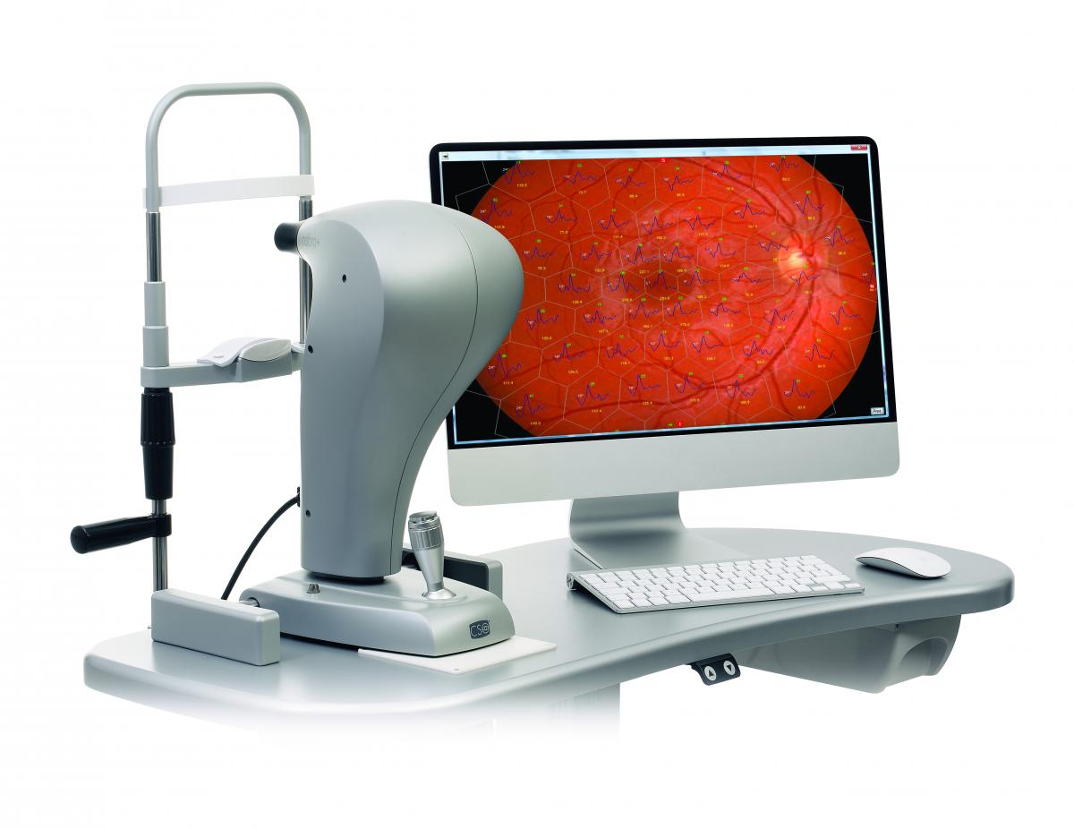
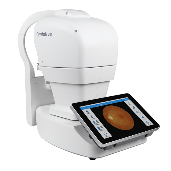
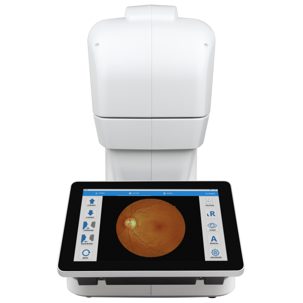
CSO Combra+ Fundus Camera is an easy to use, non-mydriatic digital fundus camera. The 5 mega pixel camera produces high resolution retinal images up to a 50 degree visual field. Cobra+ can capture images through a very small pupil diameter (2.5mm).
Images can be transferred to the Phoenix software platform for analysis and review. Cobra+ has 9 internal fixation points which allows the capture of the peripheral retina, in order to give a panoramic image of the peripheral areas. The automatic measure of the “Cup to Disk” ratio is very useful and fast in the glaucoma screening. Thanks to Phoenix is it possible to overlap the retinal image with the ERG multifocal test performed by Retimax.
Cobra+ is an easy to use, non-mydriatic digital fundus camera. The 5 mega pixel camera produces high resolution retinal images up to a 50 degree visual field. Cobra+ can capture images through a very small pupil diameter (2.5mm).
Images can be transferred to the Phoenix software platform for analysis and review. Cobra+ has 9 internal fixation points which allows the capture of the peripheral retina, in order to give a panoramic image of the peripheral areas. The automatic measure of the “Cup to Disk” ratio is very useful and fast in the glaucoma screening. Thanks to Phoenix is it possible to overlap the retinal image with the ERG multifocal test performed by Retimax.
-
MULTIPLE WAVE-LENGTH IMAGES
Multiple wave-length images can be displayed on one screen: the original image, infrared image red-free image; as well the choroidal, vascular and nerve fiber images. -
CUP TO DISK MEASUREMENT
The measurement of the Cup to Disk ratio is easliy achieved using the built in measurment tools that are available in the Phoenix software platform for the detection of glaucomatous disease. -
MOSAIC FUNCTION
Cobra+ can capture multiple images which can be combined together to create a panoramic image of the peripheral retina. -
INTEGRATION TOOL WITH ERG TEST*
The image of the retinal fundus provided by COBRA+ can be combined with the multifocal ERG test, perfor- med with the RETIMAX device. This new module pro- vides a precise indication of the functionality of every analyzed retinal area; it is very useful for the diagnosis and follow-up of Macular Degeneration as well as degenerative hereditary retinal diseases. *optional module -
AVR EVALUATION MODULE (Optional)
The AVR* tool measures the ratio between the branch arteriolar-venous diameter. A low ratio between the dimension of the vessels, may be predictive of cardiovascular problems in adult patients. *Available with Phoenix 3.XX
TECHNICAL DATA
| Weight: | 6Kg |
| Chin rest movement: | 70mm ± 1mm |
| Minimum height of the chin cup from table: | 24cm |
| Base movement (xyz): | 105 x 110 x 30mm |
| Working distance: | 20mm |
| LIGHT SOURCES | |
| Auxiliary | Led @850nm |
| Flash | Led @450-650nm |
| RETINOGRAPHY | |
| Spherical correction | from -15D to +15D |
| Image resolution | 2448 x 2051 (5MPixel) |
| Visual field | 50° x 45° |
| Minimum pupil size | 2.5mm |
| Internal (1 central 8 peripheral) | Interna (1 centrale 8 periferiche) |
| Compatibility with standard | UNI EN ISO 10940:2009, DICOM (IHE integration profile EYECARE Workflow) |
| REQUISITI MINIMI DI SISTEMA | |
| PC: 4 GB RAM - Scheda Video 1 GB RAM (non condivisa) risoluzione 1024 x 768 pixels - USB 3.0 type A Sistema operati vo: Windows XP, Windows 7 e Windows 10 (32/64 bit). | |
Related Products
“Sales available for the State of Florida, Caribbean, Central and South America”
With automatic 3D tracking and focusing, retinal images can be captured by single tap.
Reviews
There are no reviews yet.